Before performing any kind of radiological treatment, the factors that the radiologists should take of are: Defense from the radiation, The results of the radiations on the human body, Executing Suitably, Correct Interpretation of the test.
There is range of Radiology procedures and also every test is executed differently.
CT scan- a diet regimen is to be followed bought. The person is provided with a comparison representative by mouth, intravenously or rectally. It is a pain-free examination. In this the client is stocked a movable table as well as is glided inside a round device and also asked to hold the breath for some time. The entire process takes a time of about 15 to thirty minutes.
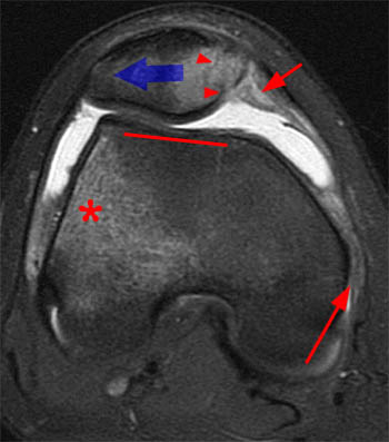
MRI scan- it is additionally a painless exam in which is positioned under a big round magnet that has a extremely high electromagnetic field. It is a safe process Radiology treatments. Some safety measures should be taken before this examination. Individuals with pace manufacturer are not allowed to go thorough this examination. The whole procedure takes around 1 to 2 hrs.
PET scan is broadened as positron emission tomography. It reveals the body metabolism as opposed to revealing the makeup. Before the test the person is carried out with contaminated sugar as well as enabled to take rest so that the sugar gets distributed thoroughly throughout the body and after that glided inside the scanner to carry on the treatment. It takes about 1.5 to 3 hours to finish.
Angiogram- in this examination a catheter is placed into the artery through which a comparison material is administered. If the tests are to be absorbed the morning after that the client is not permitted to have food as well as drink water after twelve o'clock at night. This test is a painless one and also takes close to regarding 2 hrs.
Ultrasonography- in this acoustic wave with high frequency are usage to picture inside the body and after that gotten by the transducer which is deemed an picture in the screen. USG is of numerous types. Pelvic abdominal, renal and so on. In pelvic USG the client is asked to consume 30-to 45 oz of water prior to the examination. This examination is additionally painless.
Radiology engineers take xrays and administer nonradioactive materials into individuals' bloodstreams for diagnostic purposes. Some focus on analysis imaging technologies, such as computerized tomography (CT) and magnetic resonance imaging (MRI). Radiologic engineers and also service technicians, likewise referred to as radiographers, generate xray movies (radiographs) of parts of the human body for usage in detecting medical issues.
They prepare clients for radiology tests by clarifying the procedure, eliminating articles through which xrays can not pass and placing clients to ensure that the parts of the body can be properly radiographed.To prevent unneeded radiation exposure, these workers surround the exposed area with radiation defense devices, such as lead guards, or restrict the dimension of the xray beam of light with collimation.
Radiology technologists position radiographic tools at the appropriate angle as well as height over the suitable location of a individual's body. Utilizing instruments similar to a measuring tape, they might gauge the density of the section to be radiographed as well as set controls on the xray machine to create radiographs of the proper thickness, information, and also comparison. They position the x ray film under the part of the individual's body to be taken a look at and make the exposure. They after that remove the movie and establish it.
After inspecting the movie for quality, the rad technology will send it to the radiologist for interpretation. The patient is launched as well as told to expect the results.
Radiology is an additional form of medical specialty which is utilized ti obtain pictures of different parts of the body to find as well as deal with illness. Different imaging techniques are made use family medical center of by the radiologists and also the most essential amongst them are X-RAY, USG, CT Scan, nuclear medication, PET as well as MRI.
There are various sorts of Radiology methods which are discussed as under:-.
X-Rays- it is also known as radiographs. There are created by passing x-rays via the individual's body which after that obtains directed to a catching gadget and additional developed as an image. The most frequently previously owned form of imaging is the Silver Containing movies which is now replaced by Digital radiography. As a result of its availability as well as economical prices is the most proposed examination provided by the physicians.
Fluoroscopy- Angiography or Fluoroscopy are the special form of an x-ray applications. In this a display as well as an intensifier is used which aid in the development of the image both this points are linked to a close circuit television. The client as carried out with different agents to distinguish in between the cells. It is typically made use of to identify lumps or cysts.
Interventional radiology- it is primarily made use of to identify and treat outer vascular diseases, Inferior vena cave filter positioning, gastrostomy tube placements, biliary stents and also hepatic treatments in a minimally invasive method.
Computed Tomography- X-rays is used on top of that with formulas to take image of the body. It is used for detecting immediate circumstances such as hemorrhage, embolisms in the arteries of the lungs, appendicitis, as well as treating kidney rocks.
Ultrasound-it is made use of to visualize the fetus, kidney rock, spleentomegaly etc. it made use of the high frequency acoustic wave to spot the problems.
Magnetic Resonance Imaging- it makes use of electromagnetic fields to find the nucleus of the atom within the tissues, after that makes use of a radio signals to create disturbance in the axis of rotation of center and observes the radio frequency signal generated And none the less are the nuclear medications imaging which are provided into the individuals having products which have the fondness for cells classified with contaminated tracer.
Ideally, radiology services should be available 24/7 in clinical centers for the fast interpretation of exams and also prompt therapy of clinical conditions. This is specifically crucial for emergency circumstances where time is of the essence.
Regrettably, this isn't constantly the case - especially for smaller sized hospitals, clinics or practices. The advent of teleradiology has made this possible for these institutions and also individuals, allowing them to supply faster, premium quality patient care.
Medical facility emergency rooms, medical wings, and other extremely essential clinical therapy environments frequently need radiological photos taken as soon as possible for clients who suffer from crashes or significant conditions that emerge unexpectedly. Via teleradiology, doctors have the ability to get specialist radiology solutions instantly to aid protect a diagnosis.
Specific companies are devoted exclusively to providing such solutions and use permanent radiologists who are offered round-the-clock. Such services can include a variety of specializeds and subspecialties, ranging from body imaging, to pediatric radiology, to cardiovascular imaging. Radiologists make use of the most up to date image archiving and also interactions system to receive, evaluate and translate radiological photos, such as X-rays, MRIs and CT scans.
With the ideal technology as well as high-speed Net, radiologists can execute these as well as generate both preliminary and final records from their residences, no matter the time or day. In some companies, records can even be provided in as soon as half an hour. Teleradiology has actually paved the way for a lot more quick diagnosis and also treatment.
Speed, integrity and also adaptability have actually made this practice gain raising appeal among clinical facilities across the nation. Due to the fact that when it involves person treatment, there's no time to waste.
Numerous methods are used to picture organs either directly or indirectly, Vistas of medical imaging have actually broadened explosively recently as well as are still establishing quickly.
The most time-honored and also well developed technique of imaging is radiology, which utilizes X-rays. Shadows cast on the photosensitive movie by various tissues differ in density as well as this concept is filed a claim against in interpreting the radiographs. Various techniques like simple radiography, comparison radiography as well as tomography are used. Radiological imaging provides information about physiological and also structural modifications in an organ, e.g, foreign bodies in the bronchi, loan consolidation of the lungs, cardiac enlargement, abnormalities of bones, etc. Both the anatomic abnormalities as well as physiological features can be studied by techniques utilizing comparison radiography, e.g, barium swallow, barium dish follow through, cholecystography, contrast urography, and so on. Angiography elegantly discloses the vascular supply of an body organ. Aside from envisioning occlusion and aneurysms, the vascular pattern provides indirect evidence of lumps, room occupying sores as well as additionally the practical state of the body organ. Angiography has actually been thoroughly used in Cardiovascular, neurological, renal, hepatic, as well as other disorders. Angiography has actually been made use of with various other approaches like computerized tomography to improve the resolution of information better. Angiography has actually been related to the arteries, blood vessels and lymphatics.
A new advancement in the field is interventional radiology in which investigatory or restorative procedures are done under radiological control. The technique is highly innovative, demanding very excellent ability as well as perfect group work. A couple of classical examples of interventional radiography are endoscopic retrograde cholangiopancreatography (ERCP) with elimination of pancreatic or biliary calculi; kidney artery extension through a renal artery catheter and also alleviation of coronary occlusion making use of a balloon catheter in the coronary artery.
Radiology is still the common technique of imaging since this examination answers the majority of the inquiries. In addition its global accessibility and reasonably inexpensive have actually aided to make it one of the most appropriate investigation. Though conventional readiography is noninvasive, comparison research studies are invasive in varying levels. The direct exposure to diagnostic X-rays, though little quantitatively, adds to collective irradiation gotten by the subject. It is popular that irradiation of the fetus in utero, especially during early maternity can be harmful to the infant. So additionally repeated radiographic researches can provide collective toxicity as a result of X-rays. Though the dosage and the area of direct exposure have been considerably minimized in modern devices, this danger must not be ignored and radiological research studies should be embarked on just if correctly indicated. Lots of radiological techniques are supplemented by the newer imaging strategies like Ultrasonography, isotope scanning, computerized tomography as well as nuclear magnetic resonance.